
Miniature wireless ECG, respiration and temperature monitor…A step closer to the paperless practice
Use in the exam room to diagnose cardiac abnormalities or for pre-surgical screening, surgery and recovery monitoring.
![]() VetChek Flyer
VetChek Flyer
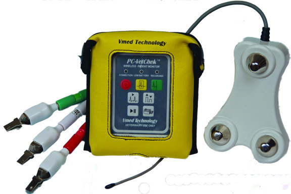
The VetChek can be used in the exam room to screen for heart abnormalities that may cause problems before, during and after surgery. ECG screens add value to the initial and subsequent annual exam and often allow the practitioner to find defects that support follow on care. Dr. Larry Tilley, a cardiology consultant, states that no other diagnostic test, including ultrasound, can accurately determine the source of various arrhythmias and conduction abnormalities as is done with ECG testing. Lead II is all that is needed in most cases; you can do away with complex lead analysis, axis shifts and perfect positioning of the pet. “The majority of veterinarians can interpret their own ECG by simply focusing on the heart rate and the cardiac rhythm.” For difficult cases, the ECG printout can be emailed directly to cardiology-based services for quick and inexpensive interpretations to determine the necessary course of action. The handy contoured chest probe makes exam room ECGs a snap. By simply placing the probe over the chest wall, the three built-in electrodes produce a quick, clean signal. The fear of the animal patient getting tangled in wires and cables is no longer an issue either. A modest procedure charge provides excellent investment payback.
ECG
“Indications for electrocardiography include arrhythmias heard on auscultation, breathing problems, shock, fainting or seizures, cardiac murmurs, and systemic disease that affects the heart (e.g. tumors, kidney dysfunction, heartworm disease). Electrocardiography is also useful as part of the preoperative work-up in older animals, for monitoring patients during and after surgery, and for evaluating the effects of cardiac drugs. An electrocardiogram (ECG) is the only test that can accurately diagnose an arrhythmia or a conduction abnormality. And an ECG will help you decide when other diagnostic tests should be done, including blood pressure measurement, thoracic radiography, or even echocardiography.
Perform electrocardiography on a periodic basis in breeds prone to arrhythmias, especially if clinical signs are present. These breeds include boxers (myocarditis), Doberman pinschers (ventricular arrhythmias and possible cardiomyopathy), German shepherds (congenital ventricular arrhythmias), and miniature schnauzers (sick sinus syndrome and sinus arrest/block).
“It is recommended that practitioners have two electrocardiography machines: an oscilloscope and an electrocardiograph. An oscilloscope is necessary for monitoring patients during surgery, and an electrocardiograph is needed for clinical diagnostic testing. The electrocardiograph linked with a strip recorder or printer provides a permanent record. The ECG can be recorded with the patient in a standing position, or you can use a hand-held unit with the patient in any position. New wireless technology (e.g. Vmed PC-Vet—Vmed Technology, Inc.) also allows an ECG to be done without wires connected directly from an animal to the electrocardiograph.”
Larry Tilley, DVM, DACVIM (Internal Medicine)
“The majority of veterinarians can interpret their own ECG’s by just simply focusing on the heart rate and what is the actual rhythm-using just one lead such as Lead II. All the other leads are used for mean electrical axis along with the size of the complexes which sometimes can help to determine heart chamber enlargement, but usually not accurate. The best way to determine heart enlargement in animals is with a chest X-ray or echocardiogram.”
Larry Tilley, DVM, DACVIM
“ECG’s have been under-utilized in most veterinary practices. With the advent of new hand-held ECG screeners and monitors, and the simplification of interpreting results, ECG screening should become a standard component of wellness exams and pre-anesthesia procedures, just as in human medicine.”
The Business of Veterinary Medicine, Steve May, Editor
“Cardiomyopathy can be diagnosed up to 2 years before becoming clinically evident by finding VPC’s on the Lead II ECG according to a recent report.”
R.E. Whitford, DVM
“Every practice needs two ECG machines, an ECG screener in the exam room and an ECG monitor in the surgical room. No other diagnostic test can accurately determine the source of various arrhythmias and conduction abnormalities as the ECG.”
Larry Tilley, DVM, DACVIM
“I find an ECG abnormality or an audible murmur on auscultation in about 1 out of 25 seemingly healthy cats.”
Gary D. Norsworthy, DVM, DAPVP
SURGERY
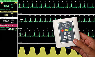
Use in surgery for ECG, temperature and respiration monitoring For surgery monitoring the PC-VetChek , when used with an optional esophageal probe, becomes a large screen patient monitor displaying digital heart rate, respiration rate and temperature and ECG and respiration waveforms. Fully programmable alarms and other settings are available in the PC-Display software. A protective hanging pouch can be mounted on the surgery table, accessory cart or IV pole. Bothersome monitoring wires and cables are no longer an issue in surgery.
RECOVERY
“The postoperative period was the most common time for dogs, cats and rabbits to die usually within three hours of surgery…greater patient monitoring and management during this time period is recommended.”
The risk of death: The Confidential Enquiry into Small Animal Perioperative Fatalities, Brodbelt, et al., Veterinary Anaesthetics and Analgesia, 2008, 35, 365-373. Post-surgical deaths.pdf